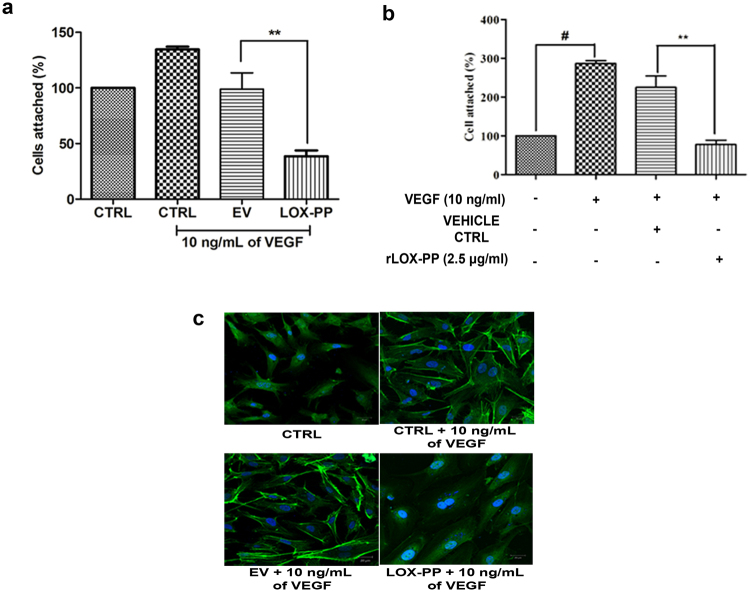
Figure 7.
Effect of LOX-PP on VEGF induced cell attachment and reorganization of F-actin: (a) Bar graph represents the quantification of cell attachment after overexpression of LOX-PP, attached cells were counted using ImageJ software (expressed as % of controls). (b) Bar graph represents the quantification of cell attachment after addition of rLOX-PP, attached cells were counted using ImageJ software (expressed as % of controls). (c) Inhibitory effect of overexpressed LOX-PP on the reorganization of actin cytoskeleton induced by VEGF (10 ng/mL) under LSM 880 confocal laser scanning microscope. HUVECs were fixed and stained for filamentous actin with phalloidin FITC conjugate. Cells were also transfected with EV. Nucleus is counter-stained with DAPI. Values were expressed as mean ± SD, n = 3. **p < 0.01 versus VEGF + EV. **p < 0.01 versus VEGF + Vehicle ctrl. #p < 0.05 versus CTRL. (CTRL – Control; EV – Empty vector; Vehicle ctrl – Vehicle control).

