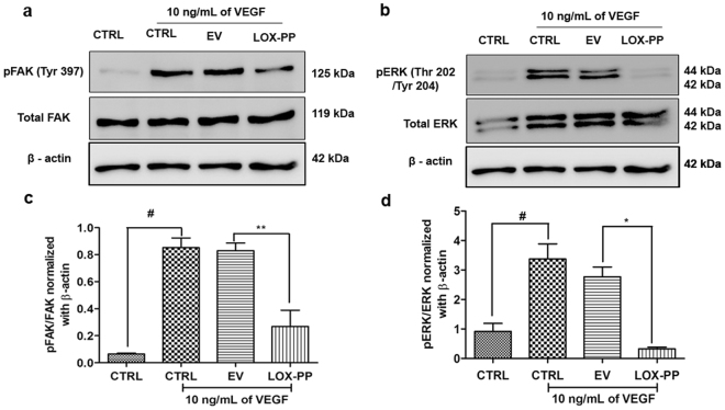
Figure 9.
Inhibitory effect of LOX-PP on FAK (Tyr397) and ERK (Thr202/Tyr204) phosphorylation induced by VEGF in HUVECs: (a) HUVECs were transfected with LOX-PP and EV. After transfection, cells were treated with 10 ng/mL of VEGF for 15 min. Whole cell extract were prepared and the samples were subjected to western blotting using antibodies against pFAK (Tyr397), total FAK and β–actin was used as loading control. The full-length blots are represented in Supplementary Figs 7, 8 and 11. (b) HUVECs were transfected with LOX-PP and EV. After transfection, cells were treated with 10 ng/mL of VEGF for 15 min. Whole cell extract were prepared and the samples were subjected to western blotting using antibodies against pERK (Thr202/Tyr204), total ERK and β–actin was used as loading control. The full-length blots are represented in Supplementary Figs 9, 10 and 11. (c) Bar graph represents quantification of the western blot using ImageJ software for FAK (Tyr397). (d) Bar graph represents quantification of the western blot using ImageJ software for ERK (Thr202/Tyr204). Values were expressed as mean ± SD, n = 3. **p < 0.01, *p < 0.05 versus VEGF + EV. #p < 0.05 versus CTRL. (CTRL – Control; EV – Empty vector).

