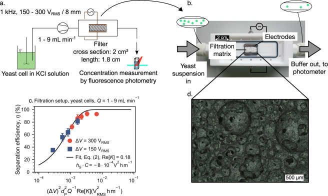
Figure 4.
Separation of baker’s yeast cells using a macroscopic porous medium (a and b). The separation efficiency η plotted against can be fitted by Eq. (2) using C⋅hS as fitting parameters (c). Experiments were conducted using two different applied voltages ΔV = 150 VRMS and 300 VRMS and throughputs in the range of Q = 60–540 mLh−1. The employed mullite open cell foam had a fluidic cross-sectional area of 2 cm2, a volume based porosity of 83% and a d50,3 of 230 μm (panel d shows an optical microscopy image of a filter cross section).

