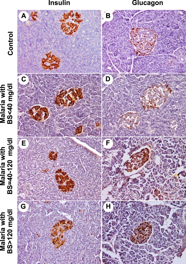
Figure 3.
The immunohistochemical staining for insulin and glucagon. Representative sections of human pancreas immunostained for insulin (left panel) and glucagon (right panel) from normal pancreatic tissues (A and B), pancreatic tissues of P. falciparum malaria patient with BS < 40 mg/dl (C and D), pancreatic tissues with BS = 40–120 mg/dl (E and F) and pancreatic tissues with BS > 120 mg/dl (G and H). All images are ×200 magnification.

