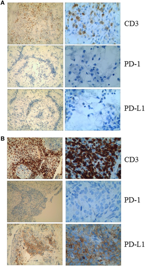
Figure 2.

(A) Immunohistochemical analysis in Patient 1 shows no membranous expression of programmed cell death-1 or PD-L1 in the tumor cells and the small background cells surrounding the malignant tumor cells. CD3+ cells are observed among the tumor cells. (B) Immunohistochemical analysis of a pretreatment tumor in Patient 2 shows focal membranous expression of PD-L1 in the tumor cells, with additional expression seen on the small background cells surrounding the malignant tumor cells. Abundant CD3+ cells are identified among the tumor cells (magnification: left panel, 10×; right panel, 40×).
