Figure 5.

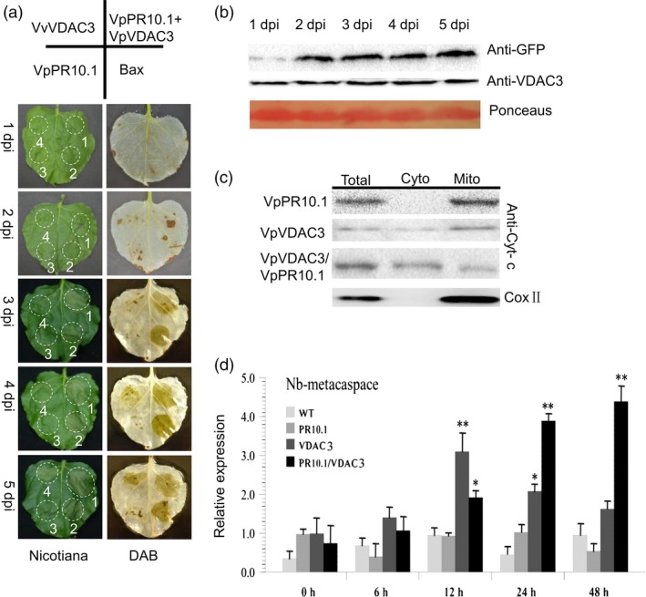
Transient Expression of VpVDAC3 or VpVDAC3/VpPR10.1 in Nicotiana benthamiana induces cell death. Phenotypic and physiological analyses of VpPR10.1 and VpVDAC3 following DAB staining. (a) Phenotypes of VpVDAC3, VpPR10.1, VpVDAC3/VpPR10.1 and Bax were all injected into one N. Benthamiana leaves. The bluish colour of DAB staining represents accumulated H2O2. (b) Visualization of VpVDAC3, VpPR10.1 protein by Western blotting in Agro‐infiltration N. benthamiana leaves. Ponceaus staining stand for control loading. (c) Western blot for detecting cytochrome c after expression with indicator gene in N. Benthamiana leaves. Total, nonseparated protein; Cyto, nonmitochondrial protein; Mito, mitochondrial enriched protein. COX II was present as mitochondrial marker protein. (d) Quantification of RT‐PCR analysis of cell death inducing gene Nb‐MCA1 expression. Values mean ± SE (n = 3) of three independent biological repeats. Asterisks indicate significant difference between each gene and GFP control, *P < 0.05; **P < 0.01 (t‐test).
