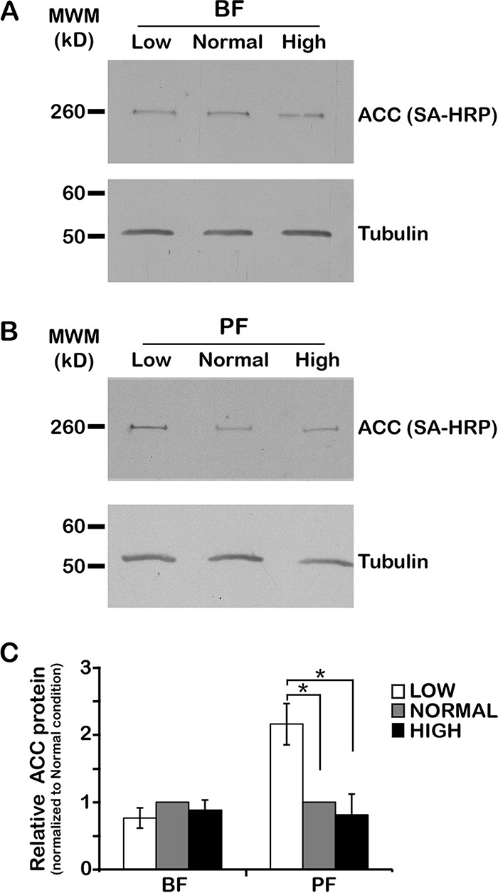
FIG 1 .

Environmental lipids affect TbACC protein levels in PF but not BF. (A and B) BF (A) and PF (B) wild-type (WT) cells were grown in low-, normal-, and high-lipid media to the mid-logarithmic (mid-log) phase (~3 days). Lysates prepared in the presence of a phosphatase inhibitor cocktail were resolved by the use of SDS-PAGE (10 µg total protein/lane) and transferred to nitrocellulose. TbACC was detected by SA-HRP blotting (A and B, top panels), and the same blots were reprobed for tubulin as a loading control (bottom panels). Representative blots from three independent experiments are shown. MWM, molecular weight marker. (C) Densitometric quantification of the results from the three independent experiments described in the panel A and B legends. The TbACC signal was normalized to the tubulin loading control. Means ± standard errors of the means (SEM) are shown. *, P ≤ 0.05 (two-tailed Student’s t test).
