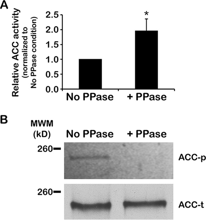
FIG 4 .

Phosphorylation of TbACC reduces activity. (A) PF TbACC-myc cells were grown in normal media to the mid-log phase (~3 days). TbACC-myc was immunoprecipitated from lysates and directly treated on-bead with 400 U of Lambda phosphatase (+PPase) or was subjected to mock treatment as a control (No PPase). Phosphatase- and mock-treated TbACC-myc was directly assayed on-bead for ACC activity. Values were first normalized to the no-ATP negative control before averaging was performed. Average values were then expressed relative to that of normal-lipid media. Means ± SEM of results from three independent experiments are shown. *, P ≤ 0.05 (two-tailed Student’s t test). (B) To confirm dephosphorylation, results of phosphatase- and mock-treated TbACC-myc pulldown experiments were resolved by the use of SDS-PAGE and assessed for phosphorylation by phosphoprotein gel staining (upper panel, ACC-p). An identically loaded gel was prepared in parallel, transferred to nitrocellulose, and probed for total TbACC by SA-HRP blotting (lower panel, ACC-t). The image was digitally processed to enable better visualization of the bands (see Materials and Methods). This experiment was performed once.
