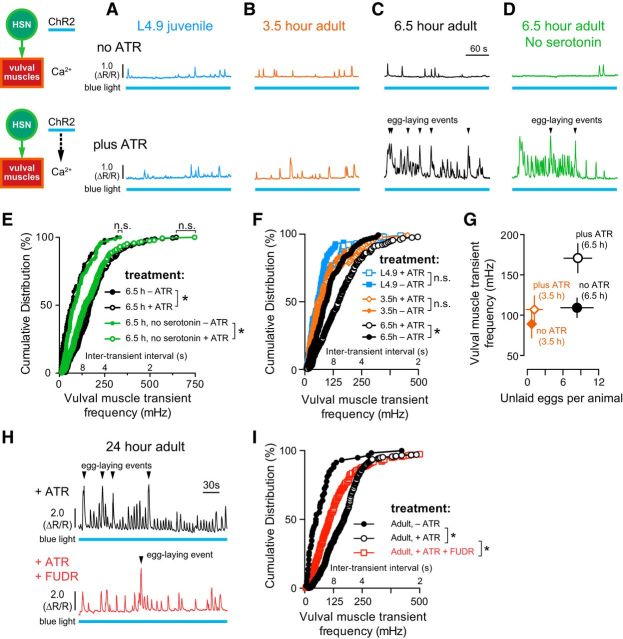
Figure 6.
Vulval muscle responsiveness to HSN input correlates with egg accumulation. A–D, Representative traces of vulval muscle Ca2+ activity in L4.9 juveniles (A, blue), 3.5 h adults (B, orange), 6.5 h wild-type adults (C, black), and 6.5 h serotonin-deficient tph-1(mg280) mutant adults (D, green) with and without optogenetic activation of HSN. Animals were grown in the presence (+ATR, top) or absence (–ATR, bottom) of ATR (see diagram). Continuous 489 nm laser light was used to stimulate HSN ChR2 activity and excite GCaMP5 fluorescence simultaneously for the entire recording. Arrowheads indicate egg-laying events. Blue bars under the Ca2+ traces indicate the period of continuous blue light exposure. E, Cumulative distribution plots of instantaneous peak frequencies (and intertransient intervals) of vulval muscle Ca2+ activity in 6.5 h adult wild-type (black filled circles, no ATR; black open circles, plus ATR) and tph-1(mg280) mutant animals (green filled circles, no ATR; green open circles, plus ATR). *p < 0.0001; n.s. indicates p ≥ 0.2863, Kruskal–Wallis test. F, Cumulative distribution plots of instantaneous peak frequencies (and intertransient intervals) of vulval muscle Ca2+ activity in L4.9 juveniles (blue filled squares, no ATR; blue open squares, plus ATR), 3.5 h adults (orange filled circles, no ATR; orange open circles, plus ATR), and 6.5 h adults (black filled circles, no ATR; black open circles, plus ATR). *p < 0.0001; n.s. indicates p ≥ 0.3836, Kruskal–Wallis test. G, Plot showing the average number of unlaid eggs present in the uterus and the average vulval muscle Ca2+ transient peak frequency, ±95% confidence intervals. H, Representative traces of HSN-induced vulval muscle Ca2+ activity in untreated (top, black) and FUDR-treated 24 h adult animals (bottom, red). Arrowheads indicate egg-laying events. I, Cumulative distribution plots of instantaneous peak frequencies (and intertransient intervals) of vulval muscle Ca2+ activity after optogenetic activation of HSNs in untreated animals grown with ATR (+ATR, open black circles), FUDR-treated animals with ATR (+ATR, open red circles), and in untreated animals without ATR (no ATR, filled black circles). *p < 0.0001, Kruskal–Wallis test.

