
Figure 8.
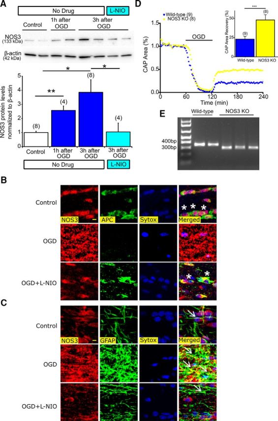
Inhibition of NOS3 upregulation in astrocytes protects oligodendrocytes and deletion of NOS3 promotes axon function against ischemia. A, MONs exposed to 60 min of OGD showed a progressive increase in NOS3 expression levels, which was attenuated with l-NIO (3 h following OGD: 3.8 ± 0.9% vs Control: 1.0 ± 0.1%), n = number of MONs. *p < 0.05, **p < 0.01; one-way ANOVA followed by Bonferonni's post hoc test. B, Colocalization of APC(+) oligodendrocytes, NOS3 (red), and Sytox (blue, nuclear staining) showed upregulation of NOS3 with OGD and loss of oligodendrocytes (white asterisks, right, merged). l-NIO (1 μm) application attenuated NOS3 upregulation and preserved oligodendrocytes. Scale bar, 5 μm. C, Colocalization of GFAP(+) astrocytes, NOS3 (red), and Sytox (blue, nuclear staining) showed upregulation of NOS3 mostly colocalized to activated astrocytic soma and processes (white arrows, right, merged). l-NIO (1 μm) application attenuated NOS3 upregulation in astrocytes and preserved oligodendrocytes. D, Axon function in MONs obtained from NOS3 knock-out mice showed improved axon function recovery following 60 min of OGD compared with control (WT) mice. n = number of MONs. ***p < 0.001; unpaired Student's two-tailed t test. E, Genotyping confirmation results for amplified DNA from tail samples. Note that the mutant band (NOS3−/−) was observed at 330 bp and the WT band (NOS3+/+) was observed at 337 bp on a 1.5% agarose gel.