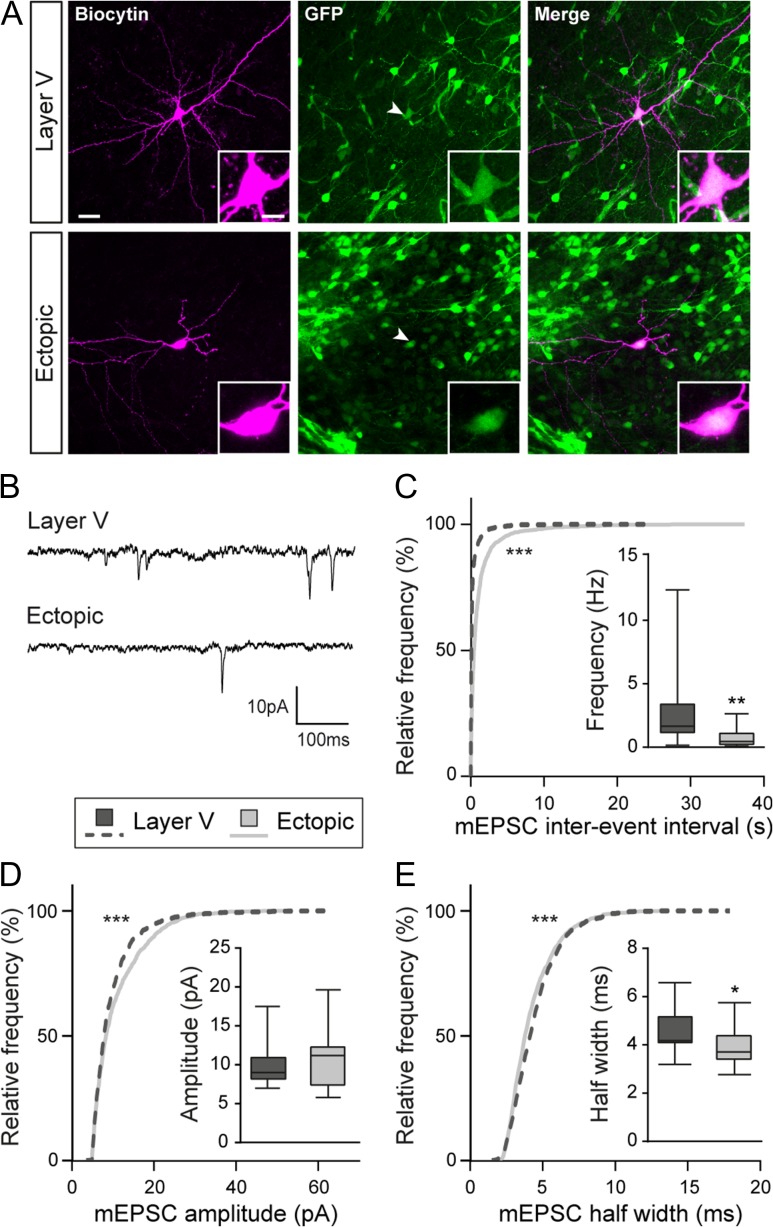
Figure 4.
Miniature glutamatergic currents are impaired in ectopic neurons. (A) Confocal images of layer V and ectopic neurons filled with biocytin during electrophysiological recordings. Recorded cells were electroporated neurons as indicated by their GFP+ fluorescence (arrow heads). Inserts show higher magnification images of the somas. Scale bars = 30 μm/10 μm. (B) Representative traces of the miniature excitatory postsynaptic currents (mEPSCs) recorded from layer V and ectopic neurons and analyzed in (C–E). (C–E) Cumulative distributions and box plots of the inter-event interval (C), amplitude (D), and half width (E) values of mEPSCs from layer V and ectopic neurons. Box limits indicate the 25th and 75th percentiles; whiskers extend to minimum and maximum values; horizontal lines show the medians. Kolmogorov–Smirnov test, P-value < 0.0001; Mann–Whitney test, (C) P-value = 0.0026, (D) P-value = 0.5053, (E) P-value = 0.0362.

