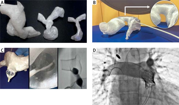
Figure 1.
A – Variability of RVOT – homograft – pulmonary branches region illustrated by three 3D printed models of (from left to right) 34-year-old, 12-year-old and 8-year-old subjects, top-down presentation. B – Model of the treated patient in the cath-lab, right lateral presentation best showing the narrowing of the homograft. C – Procedure simulation using the model – outside, inside and fluoroscopy view. D – Image documenting excellent procedure outcome: proper placement of the Melody valve and no regurgitation

