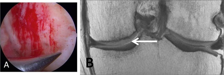
Figure 2.
A 46-year-old male laborer who had surgical repair of a high-grade cartilage defect by a microfracture technique. (A) Microfracture or perforation of the subchondral bone. Note the filling of the defect with blood clot. (B) Coronal proton density-weighted TSE MRI at 10 months post microfracture treatment demonstrates new growth of fibrocartilaginous repair tissue over the defect.

