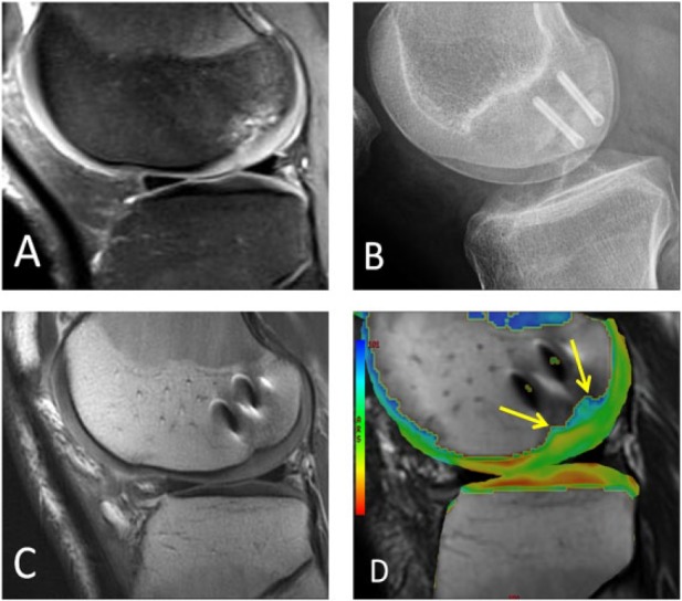
Figure 8.

A different male patient who underwent open reduction and internal fixation of a large osteochondral lesion. (A) Large loose OCD lesion is seen on sagittal fat-suppressed proton density-weighted MRI in the lateral femoral condyle. (B) Postoperative lateral radiograph shows two metallic screws fusing the OCD lesion. (C) Postoperative sagittal proton density-weighted MRI shows integration of the fused OCD lesion with susceptibility artifact from screws. (D) Postoperative sagittal T2 mapping MRI shows prolongation of relaxation times with a lack of hyaline orientation over the central weight-bearing surface of the condyle over the lesion (arrows).
