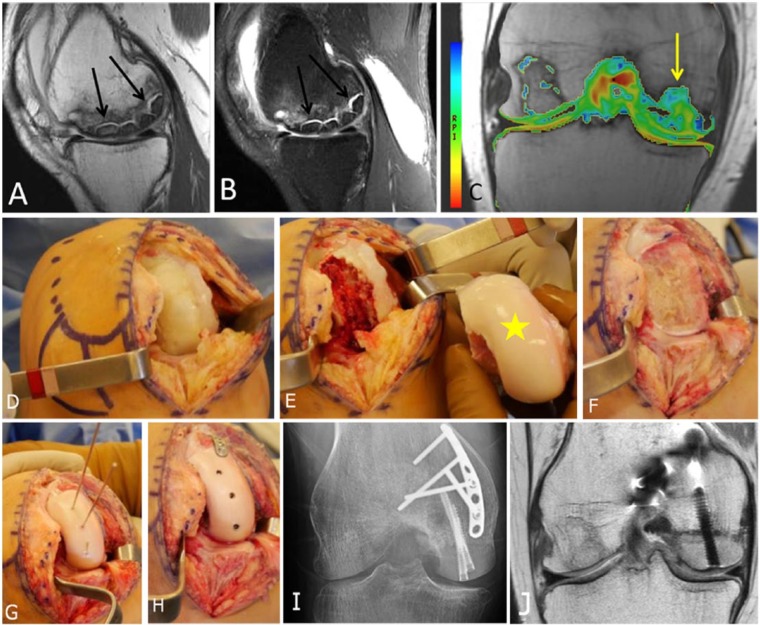
Figure 9.
A 26-year-old female who had medial femoral condyle transplantation with matched fresh allograft. (A, B) Large areas of avascular necrosis is seen in both the sagittal (A) T1- and (B) fat-suppressed T2-weighted MRI (arrows). (C) Coronal T2 mapping MRI also shows prolongation of tissue relaxation times in the cartilage over the area of transplanted medial femoral condyle (arrow) compared to the lateral condyle. (D) Open arthrotomy of the knee is performed and the AVN of the medial femoral condyle is seen. The large area of AVN is debrided and seen in E. The matched femoral condyle is seen holding up to the defect (yellow star). (F) Free cut of the medial femoral condyle is performed. (G) The matched condyle is shaped to match the defect. Three wires are used to hold the condyle to the defect. (H) Three headless screws are inserted along with a small anti-shear plate to hold and fix the condyle transplantation. (I, J) Six months postoperative AP radiograph (I) and coronal proton density-weighted MRI (J) show metallic plate and screws transfixing the medial femoral condyle transplant in place, with evidence of successful incorporation and healing.

