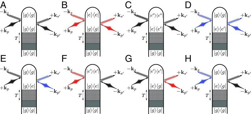
Fig. 2.
Loop diagrams for single-molecule (Eq. 4) X-ray scattering processes. The shaded area represents an arbitrary excitation that prepares the system in a superposition of and states (further explained in SI Appendix, Fig. S1). The checkered box represents a field-free propagation period that separates the state preparation from the X-ray probing process. We denote modes of the X-ray probe pulse with and , whereas , represent relevant scattering modes ( has frequency and has frequency ). Elastic scattering processes come with or and are denoted by black field arrows. Inelastic processes in which the molecule gains (Stokes) or loses (anti-Stokes) energy to the field come with or depending whether the action is on the ket or bra and are denoted with red and blue field arrows to indicate the field’s spectral shift due to the particular diagram. Note: In C, F, and G, the energy order of states is not set. We have depicted the elastic cases for specificity. Diagrams A–H identify the corresponding terms in Eq. 13.

