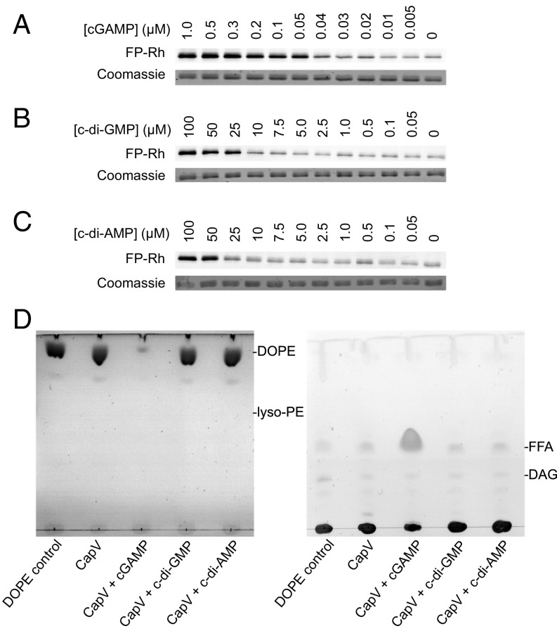
Fig. 3.
cGAMP binding directly induces CapV phospholipase activity. (A–C) Covalent labeling (Top) of the CapV active-site serine by a reactive rhodamine-labeled fluorophosphonate probe (FP-Rh). 1.6 µM His6-tagged CapV was mixed with both FP-Rh and (A) different concentrations of cGAMP or (B) c-di-GMP or (C) c-di-AMP. (Bottom) Coomassie blue staining of CapV. (D) Thin-layer chromatographic analysis of polar (Left) and neutral (Right) lipids released from the in vitro degradation of 1,2-dioleoyl-sn-glycero-3-phosphoethanolamine (DOPE) by 500 nM purified CapV in the presence of 1 µM cGAMP or other cyclic dinucleotides. DAG, diacylglyceride; FFA, free fatty acid; lyso-PE, lysophosphatidylethanolamine.

