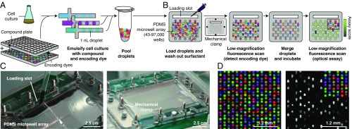
Fig. 1.
Droplets platform for combinatorial drug screening. (A) Compounds, cells, and encoding dyes are emulsified into nanoliter droplets and subsequently pooled. (B) A microwell array randomly pairs droplets (SI Appendix, Fig. S1 and Movies S1 and S2). Once loaded, free surfactant is depleted by washing to limit compound exchange. Low-magnification epifluorescence microscopy identifies the compounds carried by each droplet. Pairs of droplets in each microwell are merged and incubated (Movie S3). A second optical scan reads out a phenotypic assay (e.g., cell growth inhibition). (C) Photographs of the microwell array during loading (SI Appendix, Fig. S1 and Movies S1 and S2). Scale bars are approximate due to perspective effect. (D) Three-color fluorescence micrograph of droplets in microwell array paired with a subsequent assay of growth inhibition of E. coli cells, monitored by fluorescence from constitutively expressed GFP. Only 50% of droplet inputs contained cells; therefore, a fraction of microwells do not show GFP fluorescence. PDMS, polydimethylsiloxane.

