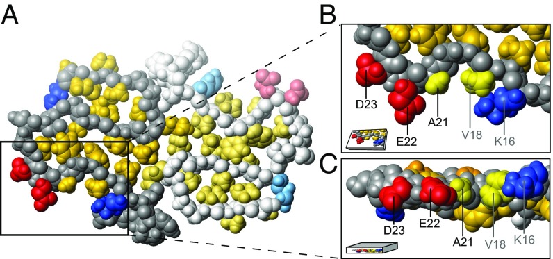
Fig. 1.
Location of residues K16, V18, A21, E22, and D23 in the A42 WT fibril model (5KK3.pdb) resolved by Colvin et al. (15). The structure within the fibril of two monomers within the same plane are shown in A, where the second monomer is displayed in paler color. The hydrophobic patch of one monomer is shown in a zoom-in top view (B) and side view (C). The image was prepared using MOLMOL (16) and shows 70% of the van der Waals radius.

