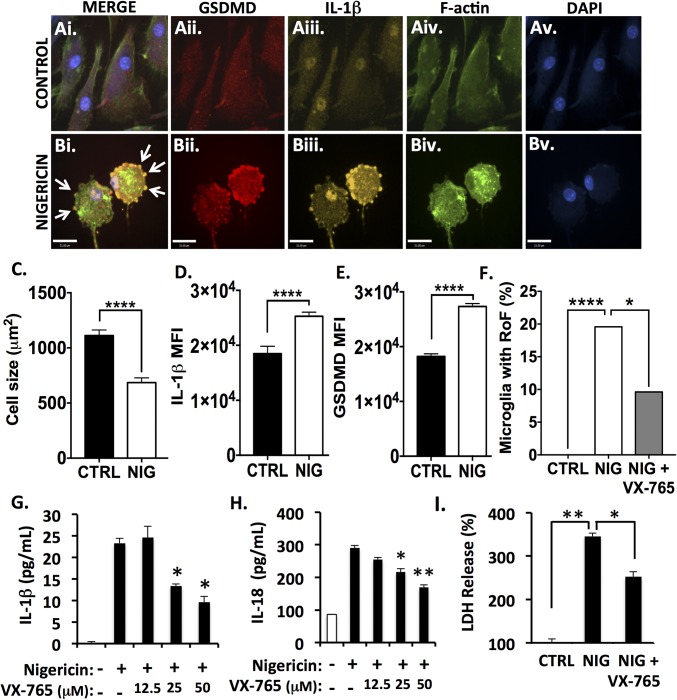
Fig. 2.
Caspase-1 inhibition by VX-765 prevents inflammasome activation and pyroptosis in nigericin-exposed human microglia. Human microglia were exposed to nigericin (5 μM, 4 h) alone or following 4 h pretreatment with VX-765 (50 μM). (A and B) Control (A) and nigericin-exposed (B) microglia were immunolabeled for GSDMD (A, ii and B, ii) and IL-1β (A, iii and B, iii) and were labeled for F-actin (A, iv and B, iv). Nigericin-exposed microglia displayed the ring-of-fire phenotype characterized by a rounded morphology and the accumulation of GSDMD+/IL-1β+ pyroptotic bodies on the plasma membrane (arrows, B, i). (Scale bars, 23 μm.) (C) Cell size was quantified for 100–150 microglia per condition (control, CTRL, or nigericin-exposed, NIG). Data represent mean ± SEM and incorporate measurements from two biological donors. (D and E) IL-1β (D) and GSDMD (E) mean fluorescence intensity (MFI) was assessed. Data represent mean ± SEM and incorporate measurements from two biological donors. (F) To quantify the formation of the ring-of-fire phenotype, 100–150 microglia per condition were classified morphologically. Data represent the percentage of microglia demonstrating the phenotype and incorporate measurements from two biological donors (χ2 test). (G and H) ELISA was used to assess IL-1β (G) and IL-18 (H) levels in supernatant from nigericin-exposed microglia. Data represent mean cytokine levels ± SEM (Student’s t test). (I) Loss of cell membrane integrity was assessed using an LDH activity assay (Student’s t test). LDH and ELISAs were performed with technical replicates of three to six wells per condition, and data were replicated in a minimum of three donor samples. *P < 0.05, **P < 0.01, ****P < 0.0001.

