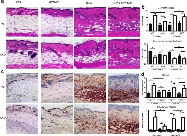
Fig. 4.
Protection from fibrosis in adipocyte nuclear corepressor-knockout (AKO) mice is mediated by peroxisome proliferator activated receptor-γ (PPAR-γ). Twenty- to 30-week-old AKO and wild-type (WT) littermate control mice were administered PBS or bleomycin (BLM) for 14 days, and lesional skin was harvested for analysis. The selective PPAR-γ inhibitor GW9662 (Sigma-Aldrich) was coadministered by intraperitoneal injection for 21 days. Injections were given as previously described (Refs [19] and [45]) as daily is not quite accurate. a Inhibition of PPAR-γ reversed the nuclear corepressor’s antifibrotic effect. H&E stain; representative images are shown. Scale bars = 100 μm. b Quantification of dermal (top) and intradermal adipose layer thickness (bottom). Results represent fold changes relative to control mice in two three mice per treatment condition and five high-power field areas per mouse; mean ± SD. * p ≤ 0.05 by analysis of variance. Results were consistent across two different experiments. c IHC was performed for macrophages; F4/80 staining is shown. d Relative F4/80 staining intensity in the dermis and intradermal adipose tissue was determined in two randomly chosen regions using Fiji software. Results represent mean ± SEM. * p < 0.05

