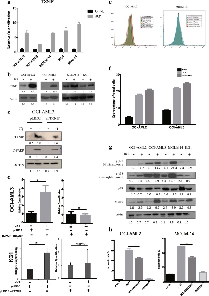
Fig. 2.
TXNIP partially mediates the JQ1-induced apoptosis through activating the p38 MAPK pathway instead of increasing ROS. TXNIP (a) mRNA and (b) protein level after JQ1 treatment. Cells were treated with JQ1 at respective IC50 for 24 h before being harvested and lysed. For real time qPCR results, means and standard errors were shown. c OCI-AML-3 cells were transfected with either pLKO.1 or pLKO.1-shTXNIP vectors and treated with JQ1 or DMSO for 24 h. d Relative quantification of apoptotic cells percentage using flow cytometry. Triplicates were performed and standard errors were shown. Percentages of apoptotic cells were normalized to DMSO-treated control. e Flow cytometry results showing the ROS level change in OCI-AML2 and MOLM-14 cells. Cells were treated with JQ1 at respective IC50 and harvested at different time points. f Percentages of apoptotic cells measured by flow cytometry. Cells were pre-treated with NAC for 1 h before adding JQ1. g AML cells were treated with JQ1 for 24 h and subjected to immunoblot analysis with primary antibodies indicated. h p38 MAPK inhibitor rescued cells from JQ1-induced apoptosis. Percentages of apoptotic cells were shown (*: p < 0.05, **: p < 0.01)

