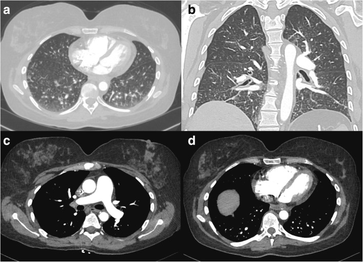
Fig. 2.
Contrast enhanced CT image with axial (a) and coronal (b) sections demonstrating a prominence of interstitial markings predominantly in posterior and basilar lung fields. In mediastinal windowing accentuating the pulmonary vasculature (c) the pulmonary artery trunk is enlarged and (d) the right ventricle and right atria appear larger in area than their corresponding left sided chambers; findings suggestive of pulmonary hypertension

