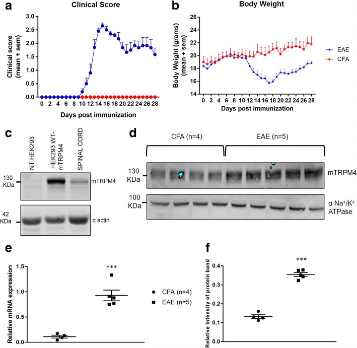
Fig. 1.
TRPM4 expression analysis in spinal cords from EAE mice. Female C57BL/6 N WT mice were immunized with MOG35–55 peptide or with CFA only. Clinical score (a) and body weight (b) were measured for 28 days after immunization. At day 28 post immunization, spinal cords were extracted and membrane proteins were isolated. c Samples from HEK 293 cells transiently transfected with mouse TRPM4 WT plasmid or empty vector (pcDNA4TO) and a spinal cord sample from a healthy WT C57BL/6 N were analyzed for TRPM4 expression, using alpha actin as loading control. A TRPM4 band can be detected at 134 kDa. d TRPM4 membrane expression was analyzed with Western Blot, and alpha subunit of Na+/K+ ATPase is used as a loading control and for TRPM4 normalization (e) qPCR analysis on Trpm4 gene expression in spinal cords from EAE mice and healthy controls. Data are represented as relative mRNA expression and the expression of Gapdh is used as reference. f Data have been analyzed using unpaired Student’s t-test and are represented as mean ± s.e.m. (*** P < 0.001)

