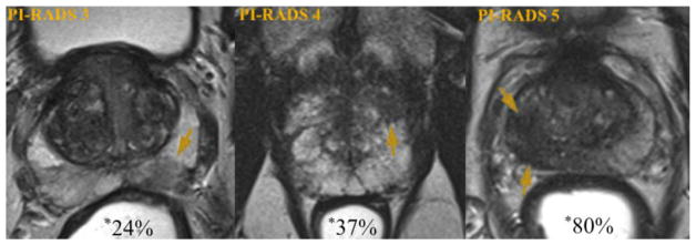
Fig. 1.
Likelihood of significant prostate cancer detection by MRI suspicion. Axial T2-weighted prostate images demonstrating peripheral zone lesions (arrows) of increasing suspicion. Examples are of a PI-RADS 3 target (left), a PI-RADS 4 target (center), and a PI-RADS 5 target (right). Percentages shown with asterisk represent likelihood of targeted biopsy revealing a cancer of Gleason Score 7 or greater, based on UCLA data (n = 1,200) (78). *Modified and reprinted with permission from European Urology 69(1); Weinreb et al. [19]. Copyright 2016, with permission from Elsevier. (Color version of figure is available online.)

