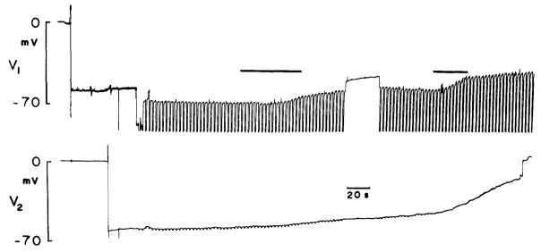
Fig. 2.
Neighboring Hensen cells impaled with single barreled electrodes, one containing KCl, the other Lucifer Yellow. Current pulses (− 5 nA) injected into Lucifer electrode and coupling responses measured in the other cell. Voltage drop across Lucifer electrode unbalanceable and thus off scale; however, since input resistance of Hensen cells is about 0.4 megaohms initial calculated coupling ratio is 0.7. Dye spred occurred in this example. Upon illumination of cells with blue light (first black bar), membrane potentials begin to fall. Subsequently, coupling response in other cell decreases. Further exposure to blue light depolarizes cells further (second black bar)

