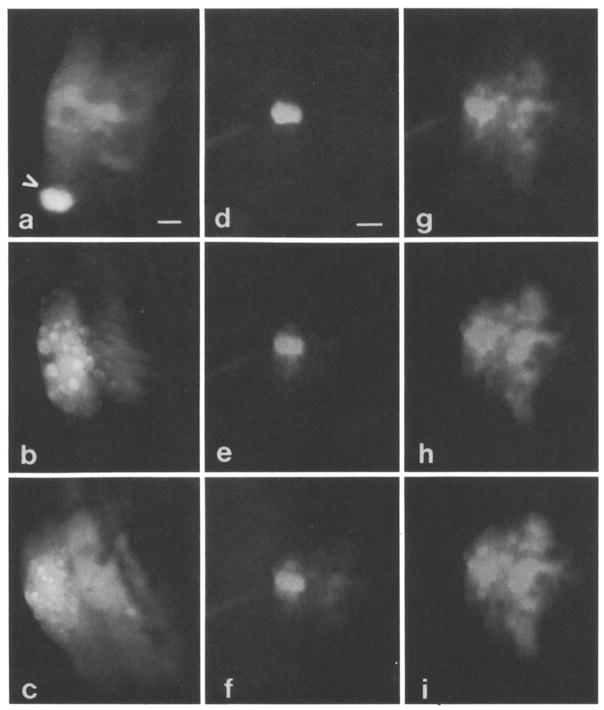
Fig. 3.
a Organ of Corti spirals around bony modiolus through which eighth nerve courses. Hensen cells are most distal to modiolus which is located to right in all photographs. Arrow depicts Hensen cell injected with Lucifer Yellow. No dye spread occurred. Subsequently, fluorescein was injected about four cells away, and dye did spread to adjacent Hensen cells as well as other supporting cells closer to modiolus. Scale: 15 μm. b Fluorescein injected into single Hensen cell, spread of dye occurred rapidly to adjacent Hensen cells and to presumed processes of Deiters cells. Bright spherical structures in Hensen cells are lipid droplets. Scale: same as in Fig. 3a. c Fluorescein observed to spread from Hensen cells to other supporting cell types of organ of Corti. All injections depicted thus far made during epi-illumination. Scale: same as in Fig. 3a. d–i Series of photographs depicting time course of spread of 6 carboxyfluorescein through supporting cells. Epi-illumination present during whole process; however, neutral density filters were used to reduce intensity. Spread of dye video taped with low light level Dage MTI camera and individual photographs taken of monitor screen, d 1 min after start of injection. Barely detectable spread in immediately adjacent Hensen cells. Scale: 15 μm for this and subsequent photographs. e 1 min 30 sec. Dye now seen clearly in spirally adjacent Hensen cells and in cells nearer modiolus, f 1 min 40 sec. Cell staining more clearly seen near modiolus, g 2 min. Spread of dye continues spirally, h 2 min 30 sec. Intensity of dye in adjacent cells approaching that of injected cell. i 3 min. Dye more uniformly distributed in group of supporting cells of different types.

