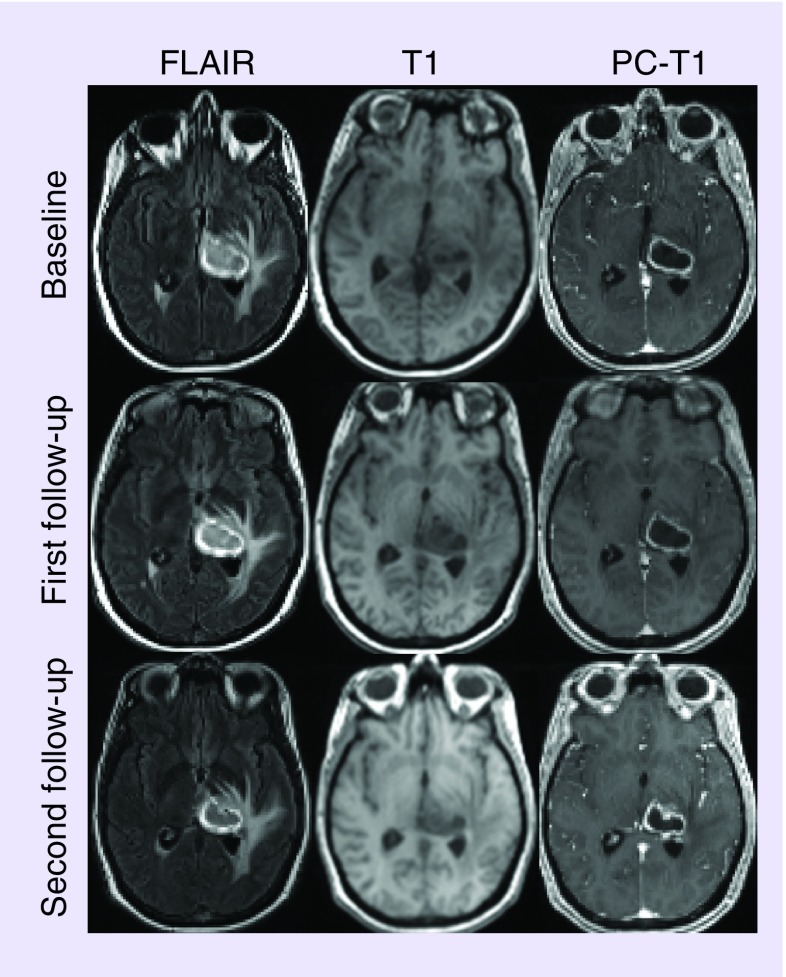
Figure 1. . A 51-year-old patient with newly diagnosed glioblastoma treated with tumor-treating fields plus temozolamide.
Axial FLAIR images at three time points demonstrate a well-defined hyperintense mass centered in the left thalamus with surrounding vasogenic edema. This mass appears as hypointense on the corresponding T1-weighted images. A heterogeneously enhancing lesion with hypointense central necrotic core and peritumoral edema is visible on the postcontrast T1-weighted images.

