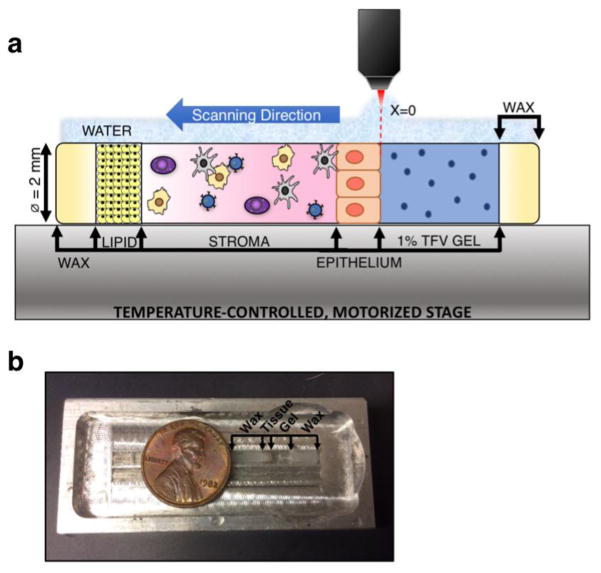
Figure 2.
a) Depiction of the lateral assay and b) aluminum holding chamber for interface with the water immersion objective; penny is shown for scale. Tissue was placed inside a 2.0 mm diameter quartz capillary tube, dosed with gel, and the ends sealed with wax. The prepared sample was then placed in an aluminum holding chamber, flooded with water, and transferred to the temperature-controlled, motorized stage for imaging with the confocal RS-OCT system. Scanning was conducted from the apical tissue surface through to the adipose tissue (lipid) layer that follows the stroma. All data points were taken at the same depth from the quartz surface. The horizontal coordinate location x=0 was defined as the tissue-gel interface.

