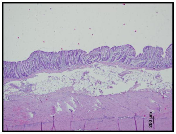
Figure 5.
Histological analysis of porcine rectal tissue (H&E stain) revealed a single-cell layer of columnar epithelium under which lay the stroma and a thick layer of adipose tissue. This corresponds well with the Raman spectra and OCT images shown in Figure 4.

