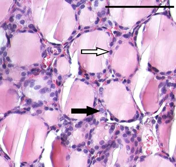
Fig 1. Microanatomy of mouse thyroid tissue.
The image shows a 20x magnification of a transverse microtome section of normal thyroid tissue from a 5-month-old female BALB/c nude mouse. Thyroid epithelial cells (follicular cells) and parafollicular cells (C-cells) are indicated exemplarily by white and black arrow, respectively. Scale bar (top right), 100μm.

