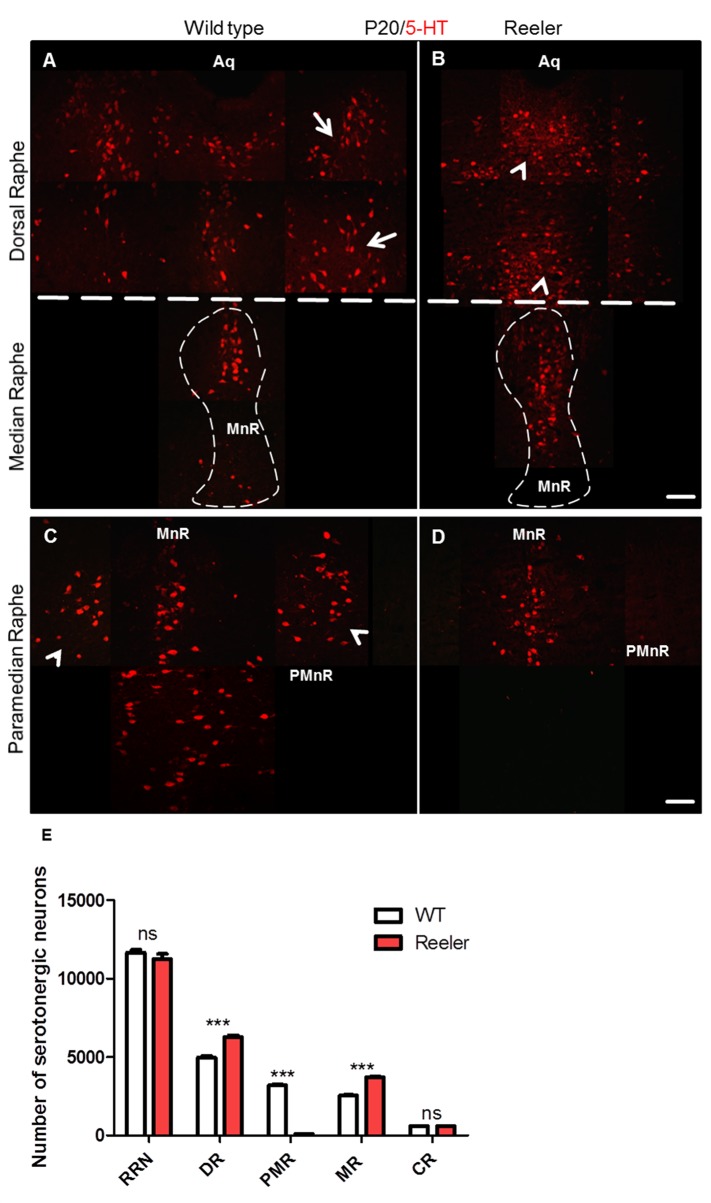
Fig 4. Loss of paramedian raphe neurons and the lateral wings of the dorsal raphe in reeler mutants at P20.
(A) Normally organized lateral wings of the dorsal raphe nuclei (white arrows) as well as median raphe in wild type mice (dashed line). (B) In reeler mutants the lateral wings of the dorsal raphe nuclei are not formed properly and 5-HT positive cells were accumulated in the dorsal raphe body (arrowheads). Moreover, relatively more 5-HT positive neurons are observed in the median raphe (dashed line) as compared with the wild type embryos. (C) Photomicrograph shows that the paramedian raphe neurons in a wild type section (arrow heads) are well organized on the both sides of the median raphe in the venrolateral region of the brainstem. (D) No formation of the paramedian raphe in reeler mutants. (E-G) Show significant reduction in the number of 5-HT- positive neurons of paramedian raphe and in turn higher neuron numbers in the median as well as dorsal raphe neurons in reeler mutants as compared to wild type mice. Thus, the total number of rostral raphe neurons was not significantly different between wild type animals and reeler mutants. Data are presented as means values ± S.E.M from 4 independent animals. Differences between groups are shown ***P<0.001. Aq. Aqueduct. Scale bar A-D: 100μm.

