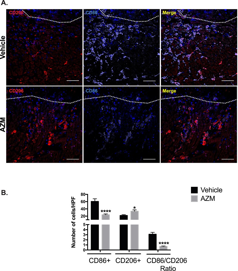
Fig 5. AZM treatment enhances alternative macrophage activation in the peri-infarct border of the injured heart.
Immunohistochemical assessment of the content of pro-inflammatory (CD86+) and reparative macrophages (CD206+) markers 3 days post-MI. Panel A shows representative images from vehicle- and AZM-treated mice demonstrating higher density of CD86+ compared to CD206+ cells in the peri-infarct border in vehicle-treated mice. White line demarcates the infarct border. Panel B shows quantitative assessment of CD86+, CD206+ and the markers ratio 3 days post-MI. The difference in pro-inflammatory and anti-inflammatory macrophages lead to a significant shift towards an anti-inflammatory state and the reduction in their ratio in AZM-treated mice (n = 4 animals/group, *P<0.05 and ****P<0.0001 compared to vehicle controls). Scale bars represent 50 μm. Data presented as mean ± SEM. AZM, azithromycin; HPF, high power field.

