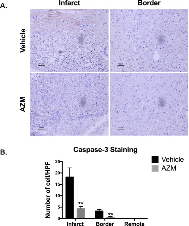
Fig 7. AZM reduces apoptosis post-infarction.
Panel A shows representative light microscope images of cleaved caspase-3 staining for infarcted and border regions in AZM- and vehicle-treated mice 3 days after MI. Quantitative analyses (Panel B) of apoptosis reveal a remarkable reduction in caspase-3 activation in the infarct and border regions of AZM-treated group compared to the control group (n = 4 animals/group, **P<0.01 compared to vehicle controls). Scale bars represent 100 μm. Data presented as mean ± SEM. AZM, azithromycin; HPF, high power field.

