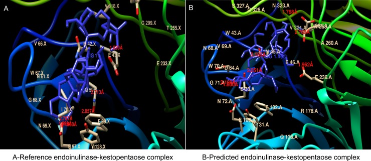
Fig 7. The Interaction between the active site around the conserved glutamate residue and substrate (kestopentaose) in the three-dimensional protein models of reference endo-inulinase from Aspergillus ficuum and predicted endo-inulinase from Talaromyces sp.
The docking was performed using MGL python tool and autodock 4. The interactions and hydrogen bonds formed between the substrate and the active site of enzyme was visualized using UCSF Chimera. The hydrogen bonds formed and their size were indicated in red color.

