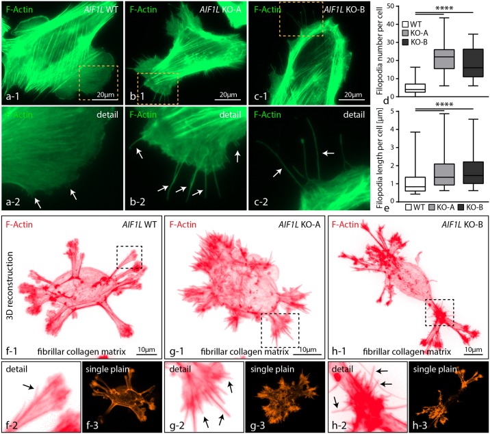
Fig 4. AIF1L prevents formation of filopodial extensions in podocytes.
(a-c) Immunofluorescence studies revealed the presence of numerous thin filopodial extensions evolving prom pre-existing lamellipodial structures in both AIF1L knockouts (dashed boxes indicate areas of higher magnification; white arrows indicate filopodial extensions). (d-e) Quantification of filopodia showed higher numbers per cell in conditions of AIF1L loss; also, measurements of filopodia demonstrated overall increased length in respective AIF1L knockout clones (n = 53 WT, 53 KO-A and 42 KO-B podocytes out of 3 independent experiments were analyzed for filopodia number; filopodia from those cells were measured for length, n = 277 WT, 1175 KO-A and 778 KO-B filopodia; **** p<0.0001). (f-h) Seeding of podocytes on thin fibrillar collagen gels for 3 hours resulted in an overall multi-polar morphology with several membrane protrusions. Numerous filopodia extended from those areas of membrane protrusion in AIF1L knockout cells. (black arrows indicate filopodial extensions; dashed boxes indicate areas of magnification).

