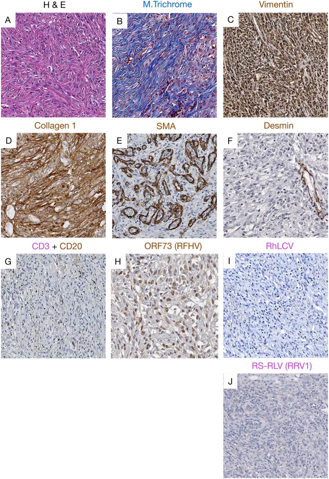
Fig 3. Representative pictures showing histopathology, immunohistochemistry and RNAscope staining of the fibrosarcoma case.
(A) H&E image showing the elongated spindle cells with euchromatin and clear nucleoli. (B) Masson’s trichrome staining showing blue collagen staining within the fibrosarcoma. Immunophenotyping demonstrated strong expression of (C) vimentin, (D) collagen I within the fibrosarcoma. (E) SMA was highly expressed in vascular smooth muscle demonstrating high vascularization of the tumor, expression of (F) desmin restricted to vascular muscles, and (G) rare expression of CD20+ B cells marker. Using IHC we detected a strong nuclear signal for (H) ORF73 proteins attesting of RFHV infection. Using next generation RNAscope approach we were not able to detect any vRNA expression of (I) RLCV or (J) RRV.

