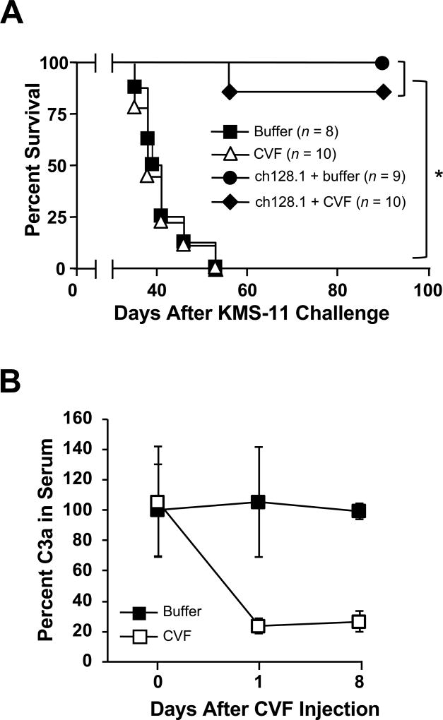
Figure 4. Role of the complement pathway in ch128.1-induced antitumor activity.
(A) Kaplan-Meier plot indicates survival of SCID-Beige mice challenged i.v. with 5 × 10⁶ KMS-11 human MM cells. Mice were injected i.v. 2 days after tumor challenge with 100 µg ch128.1. Complement depletion was achieved by injecting 25 U CVF i.p. on the day of tumor challenge and on days 3, 6, and 9. PBS was used as negative control. *p < 0.0001. (B) Confirmation of complement depletion. Blood from mice was collected before CVF injection (day 0) and on days 1 and 8. Serum C3a levels were examined by ELISA. Pooled sera collected at designated time points (n = 3–5, 1:500 dilution) were added to an ELISA plate coated with a rat anti-mouse C3a antibody. Binding was detected using biotinylated rat anti-mouse C3a, AP-conjugated streptavidin antibody and AP substrate. Values were normalized to the concentration of C3a in serum, where percent at initial time point was set at 100. Error bars show SD of samples in triplicate. Data are representative of 2 independent experiments.

