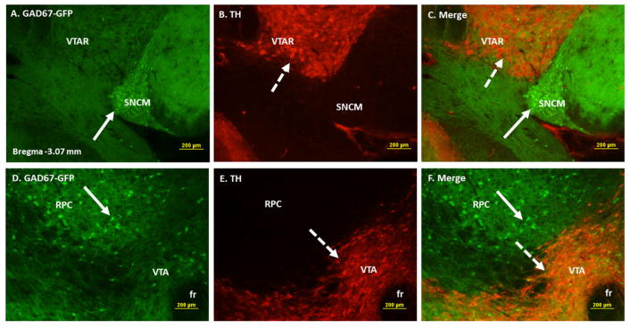
Figure 2.
Double immunofluorescence labeling demonstrating the location of GAD67-GFP positive cells with respect to the tyrosine hydroxylase (TH) immunoreactive (IR) dopaminergic neurons of ventral tegmental area (VTA). Panels A–C: High power images showing GAD67-GFP positive cells (A), TH IR cells (B) and merged images of GAD67-GFP and TH in areas overlapping VTA, SNC and SNCM. Panels D–F: High power images showing GAD67-GFP positive cells (D), TH IR cells (E) and merged images of GAD67-GFP areas overlapping VTA and RPC. GABAergic neurons are seen in close proximity to the dopaminergic neurons of VTA.
Solid arrows point to representative GAD67-GFP positive GABAergic cells and broken arrows to TH IR dopaminergic cells. Abbreviations: VTAR= ventral tegmental area rostral, SNCM= substantia nigra compact part, medial tier, fr= fasciculus retroflexus, RPC= red nucleus parvocellular part.

