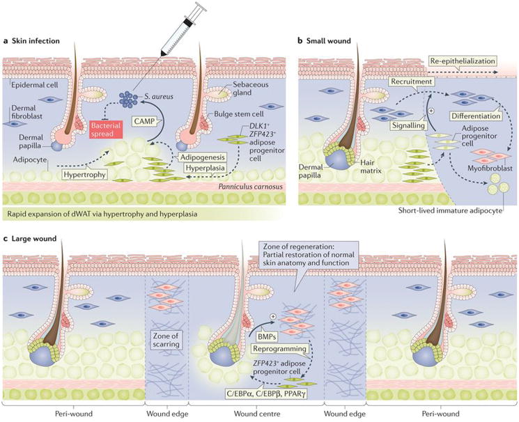
Figure 4. Role of dWAT in skin defence and regeneration in mice.

(a) Upon skin infection with Staphylococcus aureus, dWAT expands via hypertrophy and hyperplasia, irrespective of hair cycle stage. Responding to bacterial presence, expanded adipocytes secrete cathelicidin antimicrobial peptide (Camp), which has direct bacterial killing activity95. (b) Upon small excisional wounding, dWAT progenitors migrate from the wound edge into the wound bed and stimulate recruitment of fibroblasts, which are tasked with normal scar tissue formation89. (c) In large excisional wounds, new adipocytes regenerate de novo from non-adipogenic myofibroblasts. This process is driven by Bmp signals secreted by neogenic hair follicles in the wound centre. Bmps induce adipogenic lineage commitment of myofibroblasts, a process that depends on Zfp423 activation48.
