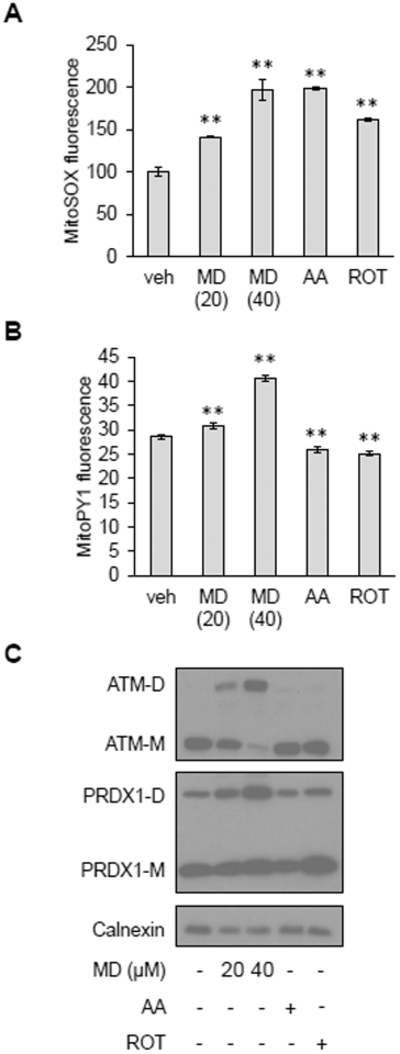
Figure 2. Conversion of mitochondrial superoxide to membrane-permeable hydrogen peroxide is necessary for ATM dimerization.

A, MitoSOX staining in HeLa cells treated with vehicle (veh), 20 or 40 μM MD, 2 μM antimycin A (AA) or 0.4 μM rotenone (ROT) for 30 minutes. Data are mean fluorescence intensities ± SD of biological triplicates. ** p<0.01 by Student’s t-test. B, MitoPY1 staining in HeLa cells treated with vehicle (veh), 20 or 40 μM MD, 2 μM antimycin A (AA) or 0.4 μM rotenone (ROT) for 30 minutes. Plotted is the mean fluorescence intensity ± SD of biological triplicates. ** as in (A). C, Western blot analysis of HeLa cells treated with vehicle (−), 20 or 40 μM MD, 2 μM antimycin A (AA) or 0.4 μM rotenone (ROT) for 30 minutes. ATM monomers (ATM-M) and dimers (ATM-D), PRDX1 monomers (PRDX1-M) and dimers (PRDX1-M) were resolved by non-reducing SDS-PAGE. Calnexin was probed as a loading control. Blots are representative of three experiments.
