. 2018 Jun 13;7:e35032. doi: 10.7554/eLife.35032

Figure 8. Schematic model showing regulation of the cell surface stability of the sheddase complex by iTAP.

(A). In WT cells the iRhom2/TACE sheddase complex successfully transits from the ER to the Golgi apparatus, where TACE undergoes maturation (prodomain removal). The sheddase complex then traffics to the cell surface, where TACE cleaves it substrates (e.g. TNF, EGFR ligands), enabling their release for signaling. iTAP, which loads onto the sheddase complex in the ER, remains associated with the sheddase complex and ensures the stability of the complex on the cell surface, promoting the cleavage of TACE substrates. (B). By contrast, in iTAP KO cells, the sheddase complex is aberrantly sorted to the lysosome, where iRhom2 and mature TACE are degraded. As a result, no TACE substrates are released for signaling. The dotted arrows indicate a putative itinerary taken by the sheddase complex in iTAP KO cells. The sheddase complex may be destabilized on the cell surface: aberrantly targeted for endocytosis and shunted to the lysosome. Alternatively, the sheddase complex may be endocytosed from the cell surface at the normal rate, but loss of iTAP may result in a defect in recycling the complex back to the cell surface, favouring delivery to the lysosome.

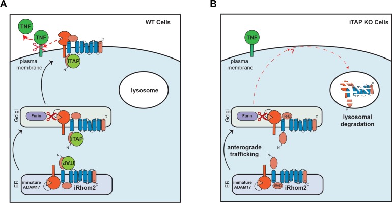
Figure 8.

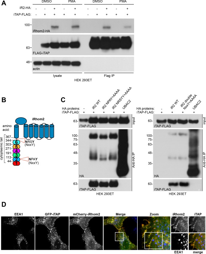
Figure 8—figure supplement 1. iTAP does not bind to features commonly recognized by FERM domain proteins.

Figure 8—figure supplement 1.

(A). iTAP appears not to have a high affinity for actin. HEK 293ET cells transiently expressing vector or iRhom2-HA and iTAP-FLAG were starved overnight then stimulated with or without PMA (1 µM) for 30 min. PMA was shown to alter the steady state of iRhom interactions with clients (Cavadas et al., 2017) and hence is used to assess potentially differential binding upon stimulated conditions. An anti-FLAG IP was performed on the cell lysates and binding was assessed by western blotting (B). Schematic representation of the location of the NxxY motifs within the iRhom2 sequence. NxxY is a common consensus binding motif for FERM proteins (e.g. sorting nexins) that participate in vesicular sorting functions. (C). iTAP binding is independent of NxxY motifs. HEK 293ET cells were transiently transfected with iTAP-FLAG and vector, iRhom2-HA, single (left hand panel) or double (right hand panel) NxxY >AAAA mutants of iRhom2-HA, or Ubac2-HA as a negative control. Cell lysates were subjected to anti-HA co-precipitation. iTAP binding to iRhom was assessed by western blotting. (D). iTAP does not co-localize with early endosomes. HeLa cells co-expressing iTAP-GFP and mCherry-iRhom2 were immunostained for EEA1, a marker of early endosomes. The white box indicates a magnified area that is separated into all three channels in the right hand images.