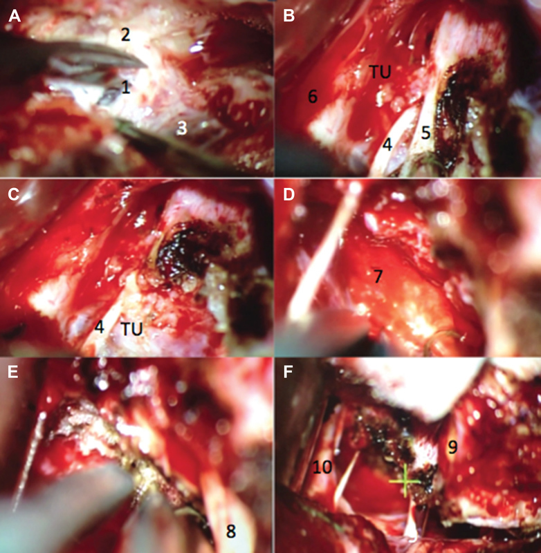
Fig. 3.

Previous case. ( A ) Presigmoid dural opening. ( B ) Coagulation of the tentorium and identification of the trochlear nerve before cutting the tentorium. ( C ) Tentorium free edge was released and reflected, noting the relationship of the tumor with the trochlear nerve. ( D ) Clivus insertion after tumor resection. ( E ) Clivus dural coagulation after tumor resection. ( F ) Cranial nerve preservation within the surgical field after tumor resection. 1. Presigmoid dura mater; 2. mastoid portion of the temporal bone; 3. sigmoid sinus; 4. trochlear nerve; 5. tentorial notch; 6. temporal lobe; 7. clivus; 8. trigeminal nerve; 9. superior petrosal vein; and 10. basilar artery. TU. Tumor.
