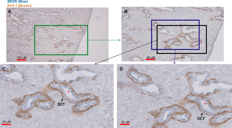
Fig. 1. BK2R is expressed in Kir4.1-positive distal tubules.

A double staining image shows the expression of Kir4.1 (brown) and BK2R (blue) with low magnification (A). Areas marked by two squares (B) are enlarged, demonstrating detailed view of BK2R staining in Fig. 1C and 1D, respectively. The distal convoluted tubules (DCT) are indicated by arrows. A red arrow indicates BK2R staining in the lateral membrane of the DCT.
