
Figure 2. Effect of EV-YF1 and CDC-exo treatment on cardiac function and hypertrophy.
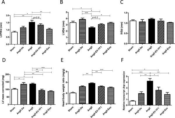
Cardiac morphology (A-D) were assessed by echocardiography at 2 weeks (AngII-2w) and 28 days after saline (sham) or Ang II infusion (AngII). Additional groups of mice were treated with Ang II plus EV-YF1 (AngII-EV-YF1) or Ang II plus CDC-exo (AngII-Exo). LVPWd: LV posterior wall thickness, end-diastole; LVIDd: LV internal diastolic diameter; IVSd: interventricular septal thickness, end-diastole. Values are means ± SEM; n = 5-10 animals/group. E, heart weight-to-body weight ratio. Values are means ± SEM; n = 7-10 animals/group. ~P<0.001 between sham and all the groups. F, relative expression of cardiac Anp by qPCR. Values are means ± SEM; n = 5 animals/group. Groups were compared using 1-way ANOVA followed by Tukey’s multiple comparisons test; *P < 0.05, **P < 0.01, ***P < 0. 001.