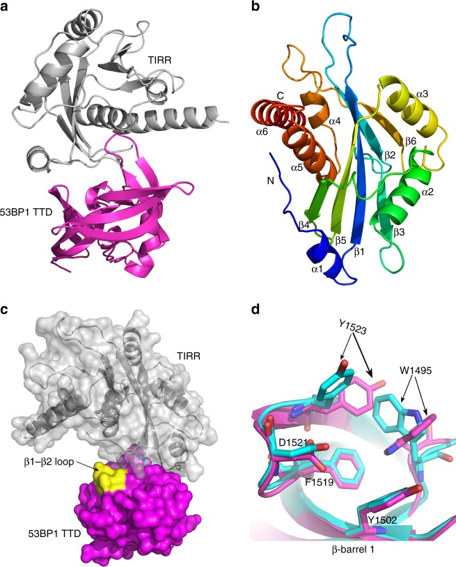
Fig. 1.
The co-structure of the TIRR–53BP1 TTD complex. a Cartoon representation of the crystal structure of the TIRR–53BP1 TTD complex. b Rainbow colored cartoon representation of TIRR. Six α-helices and six β-strands are labeled. c Molecular surface representation of the TIRR-53BP1 TTD heterodimer. TIRR is gray, 53BP1 TTD is magenta, and the subtle difference in the loop between β1 and β2 in 53BP1 TTD is displayed in yellow color. d Superposition of β-barrel 1 from 53BP1 TTD in the structure of the TIRR–53BP1 TTD complex with that of the H4K20me2–53BP1 TTD complex (PDB ID: 2IG0). The TTD in the structure of the TIRR–53BP1 TTD complex is rendered as magenta cartoon, and the TTD in the H4K20me2–53BP1 TTD complex is rendered as cyan cartoon. The amino acids corresponding to the dimethyl-lysine binding pocket are represented as stick

