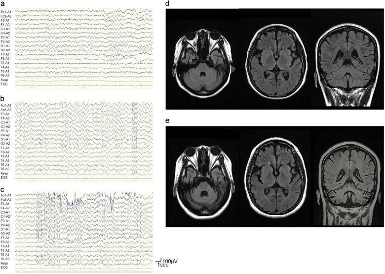
Fig. 2. Electroencephalography and magnetic resonance imaging of the proband (III-4).
a Continuous occipital slow waves were observed when she was 12 years old; b generalized spikes and slow waves, and c marked photosensitivity were observed when she was 14 years old. Brain magnetic resonance imaging (fluid attenuated inversion recovery) of the proband (III-4): d cerebral atrophy was prominent when she was 14 years old; e cerebellar atrophy appeared progressively when she was 17 years old

