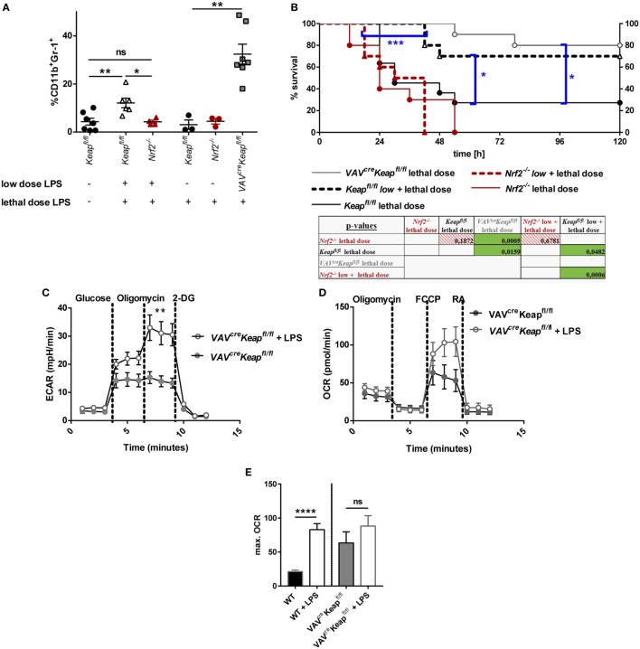
Figure 7.
Nrf2 activation in myeloid-derived suppressor cells (MDSCs) regulates LPS-mediated disease. (A) Mice were injected with either a low dose of LPS (5 mg/kg of body weight) and subsequently with a lethal dose of LPS (30 mg/kg of body weight) or solely with a lethal dose of LPS (30 mg/kg of body weight). Mice were euthanized depending on a scoring system (usually within the first 48 h after the single lethal dose) or 72 h after injection of the subsequent lethal dose and frequencies of CD11b+Gr-1+ cells were determined, unpaired, two-tailed t-test, ±SEM, N = 10 mice/group. (B) Kaplan–Meyer survival curves of mice, p-values were determined by Log-rank/Mantel-Cox Test of survival curves (single comparisons) and a subsequent FDR correction of single p-value for multiple comparison test. (C) ECAR measured under basal conditions and after addition of the indicated drugs. Points indicate mean from three control and three LPS treated VAVcreKeapfl/fl splenic MDSCs ± SEM. (D) OCR measured under basal conditions and after addition of the indicated drugs. Points indicate CD11b+Gr-1+ cells from three control and three LPS treated mice ± SEM. (E) Statistical analysis of max. OCR. Bars indicate mean ± SEM.

