Figure 1:
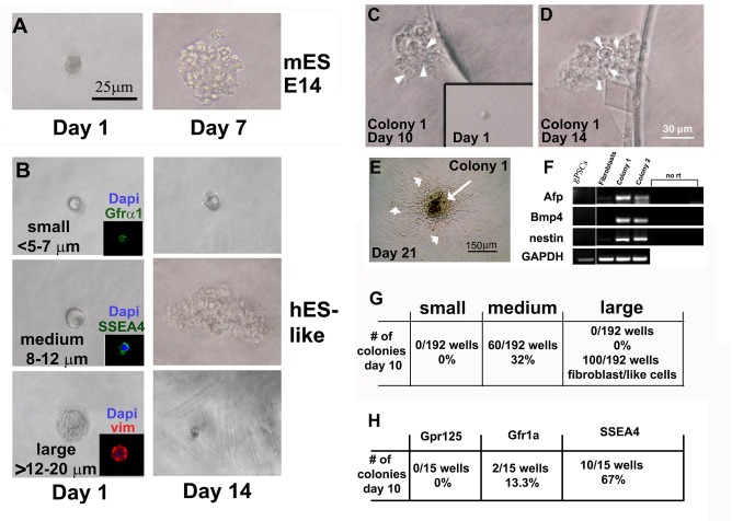
Identifying the stem cell within testes that gives rise to clonal hgPSCs. A) Acting as a positive control, a single mouse E14 strain embryonic stem cell gives rise to a colony within 7 days of culture. B) After enzymatic digestion and filtration of human testes tissue, distinctive size differences among cells were clearly evident. Small (<5-7μm), medium (~8-12μm), and large (>12-20μm) cells were isolated using a mouth pipette and placed into a well of a 96 well plate. After 14 days of incubation wells were assessed for clonal growth. Only medium size cells produced colonies. C-F) Removing bFGF from the hESC medium to induce differentiation by 10 and 14 days, respectively. Arrowheads in C and D point to individual cells within a colony, while the white circle in D outlines one cell within the colony. By ~21 days of differentiation, RT-PCR showed expression of genes from all three germ layers; Alpha fetal protein (AFP-Endoderm), Bone morphogenic protein 4 (Bmp4-mesoderm), and Nestin (neuroectoderm). However, hgPSCs did not express any of the genes from all three germ layers. GAPDH was our RT-PCR control gene. E) A central mass of differentiated cells (Arrow) are surrounded by fibroblasts, which do not show expression of these three specific genes (arrowheads in E and fibroblasts in F). G) Table quantifies colony formation from the three sizes of cells. F and insets in B) Using antibodies directed against previously explored SSC markers, SSEA4 but not Gpr125 or Gfr1α identified the cells that produced colonies of hgPSCs. SSEA4 positive cells are 8-12μm in diameter, while Gfr1α are much smaller. The large cells were mostly vimentin positive most likely representing Sertoli cells.

