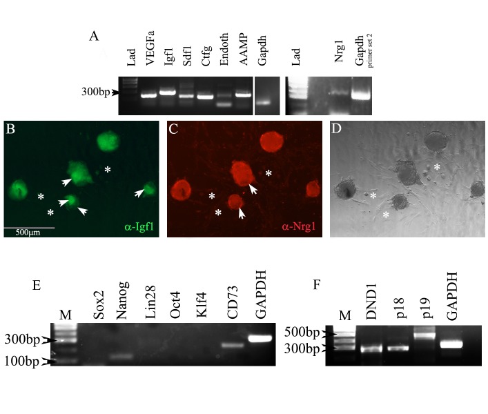
Figure 3.
Differentiation of cardiac colonies beyond day10 results in colonies that express pro-cardiac regenerative paracrine factors. A) Rt-PCR shows expression of seven pro-cardiac regenerative paracrine factors. B-D) Immunofluorescent and DIC analyses using antibodies directed against IGF-1 and NRG-1 show colonies staining positive for both paracrine factors. Arrowheads in B and C point to regions of variable staining within colonies. Asterisks highlight fibroblasts that can emanate from colonies. They show no fluorescent staining. E) Expression of most pluripotency genes is below the level of detection in differentiated colonies. Nanog and CD73 are expressed; however, nanog expression has been reported at low levels by [35] and CD73 expression has been reported as necessary for cardiac development[37]. F) Genes known to inhibit teratoma formation are expressed in hgPSC-derived cardiomyocytes. They are also expressed in hGPSCs (data not shown).

